

Diagnostic
Imaging
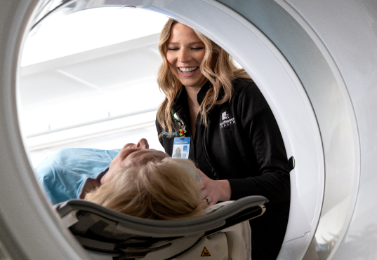
Diagnostic imaging is technology used by physicians to look inside the body for clues about a medical condition. A variety of machines and techniques create detailed images of the body’s structures and activities to help support accurate diagnosis and effective care.
At Southeast Health, we continually invest in advanced diagnostic and screening technology designed to meet the needs of both our patients and physicians. Imaging exams are performed by certified technologists and interpreted by board certified radiologists who specialize in diagnosing disease and other medical conditions.
The type of imaging your physician orders depends on your symptoms and the area of the body being examined. Most imaging tests are painless and performed efficiently with patient comfort in mind.
Once imaging studies are completed and reviewed, results are made available to patients promptly through MyChart, helping keep you informed and involved in your care.
Magnetic Resonance Imaging, or MRI, is an advanced diagnostic imaging technology that uses magnetic fields and radio waves to create highly detailed images of organs, soft tissues, bones, and joints. Because MRI does not use radiation, it is especially helpful for evaluating conditions involving the brain, spine, muscles, ligaments, and internal organs.
MRI provides exceptional detail, allowing physicians to identify abnormalities that may not be visible with other imaging exams and supporting accurate diagnosis and treatment planning.
Southeast Health is the first hospital in the region to offer advanced imaging studies using a 3 Tesla MRI, which delivers greater image clarity and shorter exam times. This higher‑strength technology allows physicians to see finer detail, enhancing diagnostic confidence.
In addition, Southeast Health also utilizes 1.5 Tesla MRI units, which are well suited for certain patient types and clinical conditions. Having access to both technologies allows our imaging team to select the most appropriate option based on individual needs, ensuring high‑quality images and patient comfort.
MRI exams are commonly used to evaluate and monitor conditions affecting the brain and spinal cord, joints, muscles, and ligaments, as well as tumors, soft tissue abnormalities, neurological conditions, and vascular or abdominal concerns.
A Computed Tomography, or CT, scan is one of the most commonly used imaging exams. It uses multiple X‑ray images taken from different angles to create detailed cross‑sectional and three‑dimensional images of the body. CT scans provide more detailed information than a standard X‑ray and are highly effective in identifying abnormalities.
CT imaging is used to support or establish a diagnosis and can help detect a wide range of conditions, including brain injuries, internal bleeding, blood clots, strokes, tumors, tissue damage, blood vessel blockages, and bone abnormalities.
CT scans are also an important tool in preventive medicine. They may be used to evaluate patients with a family history or increased risk for certain types of cancer or heart disease, helping physicians identify potential concerns early and guide appropriate care.
Ultrasound and vascular imaging use sound waves to create real‑time images of organs, soft tissues, and blood flow throughout the body. These exams help physicians diagnose conditions, guide treatment decisions, and monitor ongoing care without the use of radiation.
Ultrasound imaging is commonly used to evaluate abdominal organs, pregnancy, thyroid conditions, musculoskeletal injuries, and soft tissue concerns. Vascular imaging focuses specifically on blood vessels, helping detect blockages, narrowing, or abnormal blood flow that may contribute to stroke, heart disease, or circulatory issues.
Nuclear medicine is a specialized form of diagnostic imaging that helps physicians evaluate how organs and tissues are functioning. Instead of showing only structure, nuclear medicine exams provide insight into biological activity occurring inside the body, often allowing earlier detection of disease.
These studies use a small amount of radioactive material, called a tracer, which is absorbed by specific organs or tissues. A specialized camera then captures images that help physicians identify abnormalities and assess how well organs are working.
Nuclear medicine exams may be used to evaluate or monitor a variety of conditions, including cancer, thyroid conditions, bone disorders, infection, inflammation and heart function and flow.
Radiography, commonly known as X‑ray imaging, uses a small amount of electromagnetic radiation to create images of the body. These images, called radiographs, help physicians evaluate the type and extent of injuries such as fractures and identify disease or abnormalities in the lungs, stomach, intestines, and other areas of the body.
X‑ray images work by capturing a picture of how different tissues absorb or block radiation. Dense structures such as bones appear clearly, while softer tissues appear in varying shades of gray. Images may be recorded digitally, on specialized imaging plates, or viewed in real time using fluoroscopy.
Fluoroscopy provides continuous, real‑time X‑ray images and is often used to guide certain procedures, such as catheter placement or image‑guided treatments. This technology uses a lower radiation dose while allowing physicians to see movement and function as the procedure takes place.
A myelogram is a specialized imaging procedure performed on an outpatient basis by a radiologist to evaluate conditions affecting the spinal canal, spinal cord, and nerve roots. The exam combines real‑time X‑ray imaging, known as fluoroscopy, with computed tomography, or CT, to produce detailed images of the spine.
During the procedure, a contrast material, sometimes called X‑ray dye, is carefully injected into the spinal canal using a small needle. This contrast helps highlight the spinal structures, allowing physicians to better visualize areas of concern. The images obtained during a myelogram assist your physician in diagnosing spinal conditions and planning appropriate treatment.
Myelograms are often used when additional detail is needed or when planning for surgical treatment of the spine. These highly detailed images may help identify:
- Compression of nerve roots caused by herniated discs
- Degenerative changes in the bones or tissues surrounding the spinal canal
- Spinal stenosis
- Tumors involving the spine, spinal cord, or nerve roots
- Infections affecting the spine, discs, or surrounding tissues
- Spinal lesions caused by disease or trauma
- Cerebrospinal fluid leaks
Scheduling and Referrals
Diagnostic Imaging are performed by appointment, and most tests require an order from your physician. Our scheduling team is available to answer questions, provide preparation instructions, and help coordinate your exam.
Have Questions?
Want to learn more about volunteering at Southeast Health? We’re happy to help. Whether you’re exploring opportunities or need help getting started, we are here to guide you every step of the way.
To learn more or to schedule an appointment, talk with your provider or contact us.
Contact Us
Phone: (334) 793-8006
Once you apply, your application will be reviewed. You will receive an email or call to schedule a time for an interview to come talk with us about what you are interested in.